PET扫描
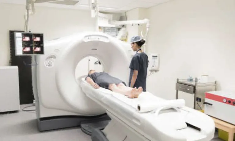
PET scanning is a gamma-based imaging technique that allows doctors to detect early signs of cancer, brain disorders and heart diseases by identifying changes in organs and tissues at cellular level. In a PET scan, the patient is injected with a radioactive substance and placed on a flat table that moves into a donut-shaped housing called gantry.
The gantry of a PET system contains several gamma ray detectors that require very precise temperature control to one another in order to generate a proper image. If one detector fails it will have a significant impact on the final image result.

正电子发射断层扫描 (PET) 是一种基于伽马射线的影像技术,它能够使医生通过在细胞水平上识别器官和组织的变化来检测癌症、脑部疾病和心脏病的早期迹象。在 PET 扫描中,患者被注射放射性物质,并被放置在一个平台,之后平台会移动到一个称为龙门架(gantry)的圆环形外壳中。
PET 系统的机架包含多个伽马射线探测器,它们都需要非常精确地控制温度以生成正确的图像。如果一个检测器出现故障,将对最终影像结果产生重大影响。
液体冷却解决方案
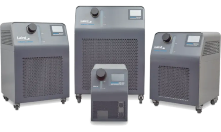
Nextreme 系列能够以 ±0.1°C 的温度精度将系统冷却至远低于环境温度,因而能够支持 PET 扫描系统的冷却需求。与以前的型号相比,Nextreme 系列是新一代循环式冷却器,噪音更小,能耗更低。
阅读相关应用笔记:
PET 和 SPECT 扫描仪的液体冷却选项
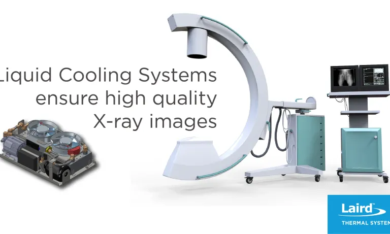
医用X射线影像设备冷却
Related Content


